What Are Human Teeth Made Of? Complete Guide to Tooth Anatomy
You use your teeth every single day. You brush them, floss them, and occasionally complain when one of them hurts. But have you ever stopped to think about what teeth actually are? What they are made of? Why they are so hard? Why they cannot heal themselves the way bone does?
Teeth are remarkable structures. They are the hardest substance in the human body, they endure thousands of pounds of force over a lifetime, and their composition is unlike anything else in your anatomy. Understanding what teeth are made of helps you appreciate why certain dental problems develop and how to prevent them.
The Four Main Tissues of a Tooth
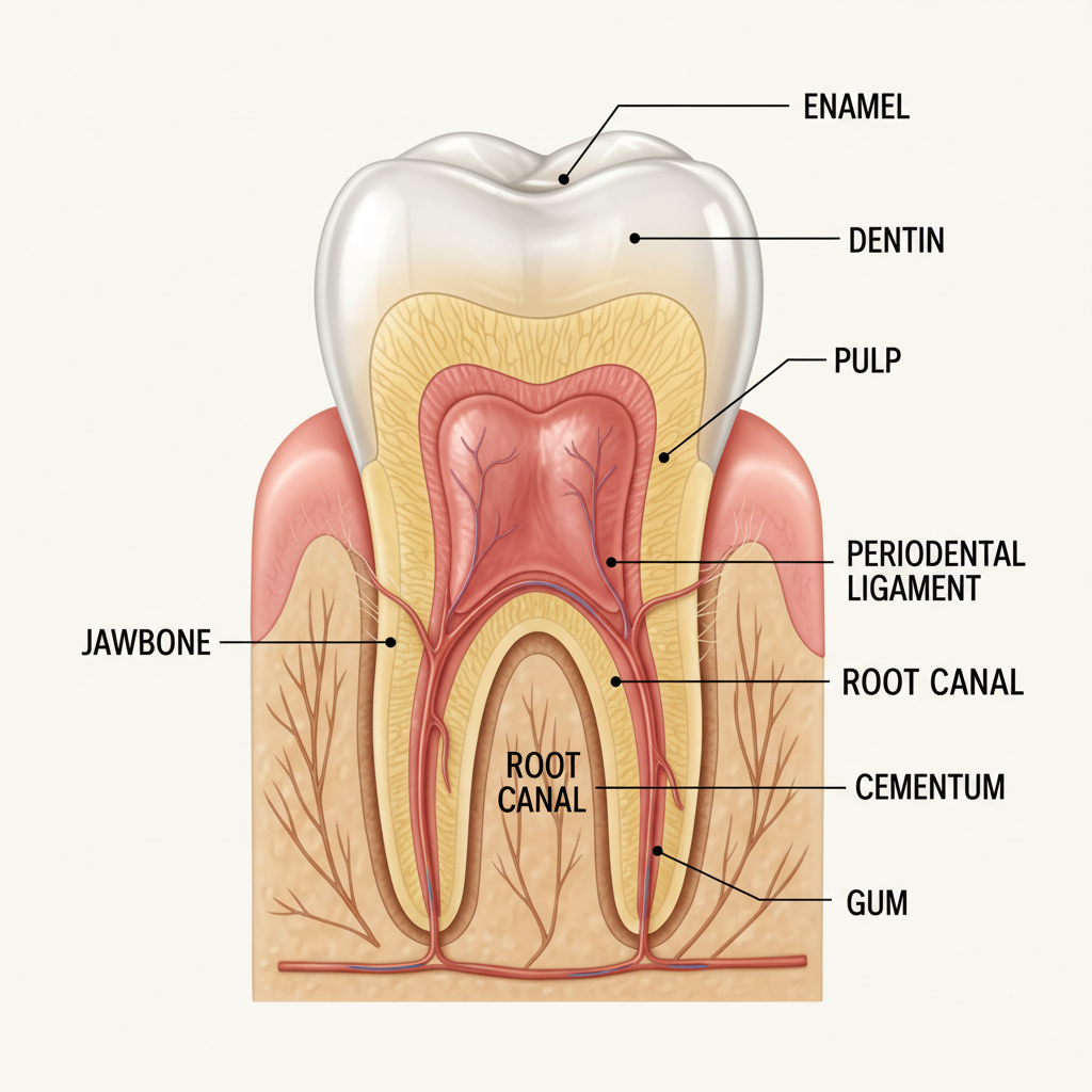
Every human tooth consists of four distinct tissues, each with a specific structure and function. From the outside in, they are: enamel, dentin, cementum, and pulp.
Enamel: The Hardest Substance in Your Body
Enamel is the outermost layer of the tooth crown, the visible part above the gum line. It is the hardest biological material in the human body, ranking 5 on the Mohs hardness scale (comparable to steel).
Composition: Enamel is approximately 96% inorganic mineral, primarily hydroxyapatite crystals (a crystalline form of calcium phosphate). The remaining 4% is water and organic proteins. This extreme mineral content is what makes enamel so hard and resistant to wear.
Structure: Enamel is organized into tightly packed rods (also called prisms) that extend from the underlying dentin to the tooth surface. Each rod is about 4 micrometers in diameter and contains millions of hydroxyapatite crystals arranged in a specific pattern. There are roughly 5 million rods in a single lower incisor and up to 12 million in an upper first molar.
Thickness: Enamel thickness varies across the tooth surface. It is thickest at the biting edges and cusp tips (up to 2.5 mm) and thins to a feathered edge at the gum line where it meets the cementum.
Key characteristic: Enamel contains no living cells. Once it is fully formed during tooth development, it cannot regenerate or repair itself. This is why cavities that penetrate enamel require a dental filling or other restoration. Your body simply cannot grow new enamel.
However, enamel can undergo remineralization, a process where calcium, phosphate, and fluoride ions from saliva and dental products are redeposited into areas of early mineral loss. This is how very early-stage decay can sometimes be reversed before a cavity forms.
Dentin: The Bulk of Your Tooth
Beneath the enamel lies dentin, which makes up the majority of the tooth structure. Dentin is softer than enamel but harder than bone.
Composition: Dentin is approximately 70% inorganic mineral (hydroxyapatite), 20% organic material (primarily type I collagen), and 10% water. The higher organic content gives dentin more flexibility than enamel, allowing it to absorb forces that might otherwise crack the brittle outer shell.
Structure: Dentin has a unique tubular structure. Millions of microscopic tubes called dentinal tubules run from the pulp chamber to the junction with enamel. These tubules are filled with fluid and cellular extensions from odontoblast cells that line the pulp. The tubular structure is why dentin is sensitive: stimuli like temperature changes, pressure, or sweet foods cause fluid movement within the tubules, which triggers nerve responses.
Types of dentin:
- Primary dentin forms during tooth development before the tooth erupts
- Secondary dentin is produced slowly throughout life, gradually reducing the size of the pulp chamber
- Tertiary (reparative) dentin is produced in response to injury or irritation, such as decay or a dental procedure. This is your tooth's defense mechanism, forming a barrier to protect the pulp
Key characteristic: Unlike enamel, dentin is a living tissue. The odontoblast cells lining the pulp continuously produce new dentin throughout your life. This is why teeth that have had deep cavities or multiple restorations often become less sensitive over time: the pulp is walling itself off by producing more dentin.
Cementum: The Root's Protective Layer
Cementum covers the root surfaces of your teeth, below the gum line. It is the interface between your tooth and the periodontal ligament that holds your tooth in its socket.
Composition: Cementum is approximately 65% inorganic mineral, 23% organic material, and 12% water. It is similar in composition to bone but lacks blood vessels and Haversian systems.
Function: The primary role of cementum is to anchor the periodontal ligament fibers that suspend your tooth in the alveolar bone. These fibers (called Sharpey's fibers) embed into the cementum on one end and into the bone on the other, creating a flexible but strong attachment.
Thickness: Cementum is thinnest at the neck of the tooth (20-50 micrometers) and thickest at the root tip (150-200 micrometers). It thickens throughout life as new layers are deposited.
Key characteristic: Cementum can be resorbed and remodeled, similar to bone. This is important in orthodontic treatment and in the body's response to chronic inflammation from gum disease.
Pulp: The Living Core
At the center of every tooth is the pulp chamber, containing the tooth's nerve, blood supply, and living cells.
Composition: The pulp is a connective tissue containing blood vessels, lymphatic vessels, nerve fibers, and various cell types including odontoblasts (dentin-producing cells), fibroblasts, immune cells, and stem cells.
Function: The pulp serves several critical roles:
- Sensory: Nerve fibers in the pulp detect temperature, pressure, and chemical stimuli. Pain from the pulp is your tooth's warning system that something is wrong.
- Nutritive: Blood vessels supply oxygen and nutrients to the living portions of the tooth.
- Formative: Odontoblast cells produce dentin throughout life.
- Defensive: Immune cells in the pulp respond to bacterial invasion, and odontoblasts produce reparative dentin to wall off threats.
Key characteristic: When the pulp becomes irreversibly inflamed or infected (usually from deep decay, cracks, or trauma), the tooth requires root canal treatment to remove the damaged tissue and save the tooth.
The Supporting Structures: What Holds Your Teeth in Place
Teeth do not sit directly in bone like posts in concrete. They are suspended by a sophisticated system of tissues collectively called the periodontium.
Alveolar Bone
The alveolar bone is the ridge of bone in both jaws that contains the tooth sockets. It is constantly remodeling in response to the forces applied through your teeth. This is the bone that resorbs when teeth are lost and the same bone that dental implants integrate with.
Periodontal Ligament (PDL)
The PDL is a thin (0.15-0.38 mm) layer of connective tissue between the tooth root and the alveolar bone. Its collagen fibers act like tiny shock absorbers, allowing slight movement of the tooth within its socket during chewing.
The PDL also contains proprioceptive nerve endings that give you the remarkable ability to detect very small objects between your teeth (you can feel a human hair) and to modulate your biting force automatically.
Gingiva (Gums)
Your gums form a protective seal around the neck of each tooth, preventing bacteria from reaching the bone and ligament below. Healthy gums are firm, pink (though pigmentation varies naturally), stippled (like orange peel), and do not bleed when brushed or flossed.
When gum tissue becomes infected through periodontal disease, the seal breaks down, allowing bacteria to migrate down along the root surface, destroying bone and ligament. This is why regular dental cleanings and exams are so important.
Types of Human Teeth and Their Functions
Adults have 32 permanent teeth (including wisdom teeth), divided into four types:
Incisors (8 total)
The four upper and four lower front teeth. They have a flat, chisel-shaped edge designed for cutting and shearing food. Incisors also play a major role in speech, particularly the sounds "th," "f," and "v."
Canines (4 total)
The pointed teeth at the corners of your dental arch. Canines have the longest roots of any teeth and are designed for gripping and tearing food. They also guide your jaw movements when you chew side to side.
Premolars (8 total)
Also called bicuspids, these sit between the canines and molars. They have two cusps (points) and a broader biting surface. Premolars are transitional teeth that both tear and crush food.
Molars (12 total, including wisdom teeth)
The large, flat teeth at the back of your mouth. Molars have four to five cusps and broad surfaces designed for grinding food into small, digestible particles. Your first molars bear more biting force than any other teeth. Wisdom teeth (third molars) often require extraction due to impaction or crowding.
How Teeth Develop
Tooth development begins remarkably early. The foundations for your baby teeth start forming around 6 weeks of gestational age, well before birth. Permanent teeth begin developing around the 20th week of pregnancy.
Enamel formation (amelogenesis) is carried out by specialized cells called ameloblasts. These cells deposit enamel matrix proteins that are gradually replaced by hydroxyapatite crystals. Once the enamel is fully mineralized and the tooth erupts, the ameloblasts die. This is why enamel cannot be regenerated: the cells responsible for making it no longer exist.
Frequently Asked Questions
Are teeth considered bones?
No. While teeth and bones share some similarities (both contain calcium phosphate minerals), they are fundamentally different. Bones are living tissue with blood vessels throughout, can heal fractures, and constantly remodel. Tooth enamel is non-living and cannot regenerate. Dentin is living but has limited self-repair capability.
What is the strongest part of a tooth?
Enamel is the strongest part, consisting of 96% mineral content. It is the hardest substance produced by any vertebrate. However, enamel is also brittle, which is why teeth can chip or crack under excessive force despite being incredibly hard.
Why can teeth not heal themselves like bones?
Bones contain osteocytes (living cells) throughout their structure and have a rich blood supply that delivers the materials needed for repair. Enamel has no living cells and no blood supply. Dentin can produce limited reparative tissue, but once enamel is lost, the body cannot replace it.
What causes teeth to turn yellow?
Tooth color is primarily determined by the color of dentin showing through the semi-translucent enamel. As enamel thins with age and wear, more of the naturally yellow dentin becomes visible. Staining from coffee, tea, wine, and tobacco also contributes. Professional teeth whitening can significantly improve tooth color.
How much force can a tooth withstand?
The average maximum bite force in humans ranges from 120 to 140 pounds (540-630 Newtons), though it can exceed 250 pounds in some individuals. Molars withstand the greatest forces. Teeth are designed to handle compressive forces well but are vulnerable to lateral or shearing forces, which is why grinding and clenching cause damage.
Do teeth have nerves?
The enamel and cementum do not contain nerves. However, the dentin has tubules connected to nerve fibers in the pulp, which is why exposed dentin is sensitive. The pulp itself contains extensive nerve networks that can produce significant pain when inflamed.
Can enamel be rebuilt artificially?
Currently, no commercial treatment can regrow natural enamel. However, dental materials like composite resin, porcelain crowns, and veneers can effectively replace lost enamel. Research into enamel regeneration using stem cells and biomimetic materials is ongoing but not yet clinically available.
---
Understanding your tooth anatomy helps you make better decisions about your oral health. At Piedmont Dental in Rock Hill, SC, we believe informed patients get better outcomes. Contact us or call (803) 328-3886 to schedule your next dental exam.